



August 29, 2026 FMX vs. CBCT: Which Dental Imaging Does Your Practice Actually Need, and When
The short answer: a full-mouth series (FMX) and a cone-beam CT (CBCT) are not two versions of the same exam. An FMX is a set of two-dimensional intraoral radiographs – the standard, everyday imaging for detecting caries, assessing bone levels and periodontal status, and building a comprehensive record. A CBCT is a three-dimensional volumetric scan, reserved for cases that genuinely have to be understood in three dimensions: implant planning, impacted or supernumerary teeth, complex endodontic anatomy, TMJ, pathology, and airway. CBCT is not simply a better FMX, and it is not a routine screening tool. The right choice is the one that answers the clinical question at the lowest reasonable radiation dose – and that decision has quiet consequences for your practice’s data, storage, and compliance long after the patient has left the chair.
Two images, two different questions
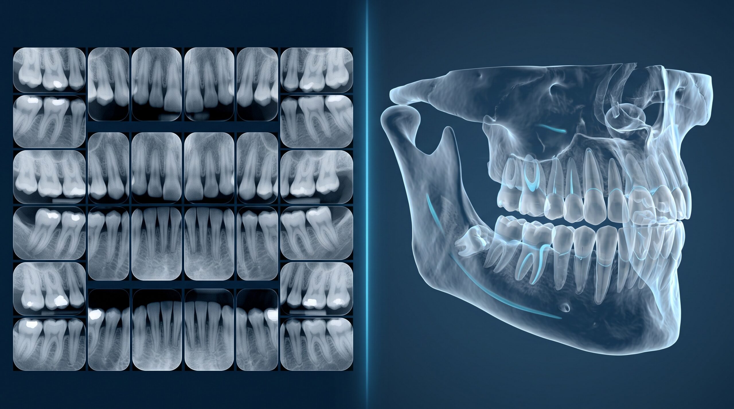
A full-mouth series is typically around 18 to 20 small intraoral images – periapicals that show whole teeth and their roots, plus bitewings that show the crowns of upper and lower teeth together. It is flat: everything the beam passes through is superimposed into a single 2D shadow. That is perfectly adequate, and appropriately conservative, for the questions general dentistry asks most often – is there decay between these teeth, how much bone is around this tooth, is this restoration intact.
A cone-beam scan is fundamentally different. It rotates a cone-shaped beam around the head and reconstructs a 3D volume you can slice in any plane and render as a model. That extra dimension is decisive when the geometry matters: how close an impacted third molar sits to the inferior alveolar nerve, whether there is enough bone width for an implant, how many canals a molar really has. What CBCT is not is a screening exam. Using a 3D scan to look for cavities is the wrong tool – more radiation, more data, and no better answer than a bitewing would have given.
FMX vs. CBCT at a glance
| Criterion | Full-mouth series (FMX) | Cone-beam CT (CBCT) |
|---|---|---|
| What it captures | 2D intraoral images of teeth and surrounding bone | 3D volumetric scan of teeth, jaws, and adjacent structures |
| Dimensionality | Two-dimensional (superimposed) | Three-dimensional (sliceable, reconstructable) |
| Typical radiation dose | Low; roughly 0.02-0.04 mSv for a digital FMX with rectangular collimation | Variable; a small field-of-view scan can approach FMX range, a large field-of-view scan is several times higher |
| Best for | Caries, periodontal/bone assessment, comprehensive records, recall | Implants, impacted teeth, endodontic anatomy, TMJ, pathology, airway, surgical planning |
| File size / data | Small – a set of modest image files | Large DICOM volume, commonly tens to hundreds of megabytes, sometimes more |
| Viewing software | Standard imaging module in your practice software | Dedicated DICOM/CBCT viewer |
| Typical frequency | Periodically, per patient risk and history | Case-by-case, only when 3D is justified |
Every row of that table is real, but the dose row deserves a caveat: CBCT dose varies enormously with the machine, the field of view, and the exposure settings, so treat these as directional, not absolute. All of these dental exams sit far below a medical CT of the same region and well within the range of everyday background radiation – which is exactly why justification, not fear, is the right frame.
Radiation, ALARA, and the justification question
Under image-selection principles endorsed by the ADA and radiology bodies – and consistent with PHIPA’s expectation that you collect only what you clinically need – you justify each exposure rather than imaging by default. That is the whole ballgame with CBCT. A 3D scan is appropriate when a 2D image would leave the clinician guessing about something that changes the treatment; it is not appropriate as a routine add-on because the machine is in the operatory and the scan is easy to take. “We have it, so we use it” is precisely the reasoning ALARA exists to prevent.
Practically, that means a documented rationale for each CBCT, using the smallest field of view that answers the question, and checking whether a recent scan already exists – a patient may have had a CBCT elsewhere that can be shared rather than repeated. This is the same discipline that governs how often you take any radiograph; we walked through the everyday version of it in our look at how many dental X-rays are too many. The modality changes; the obligation to justify does not.
The part your IT actually feels
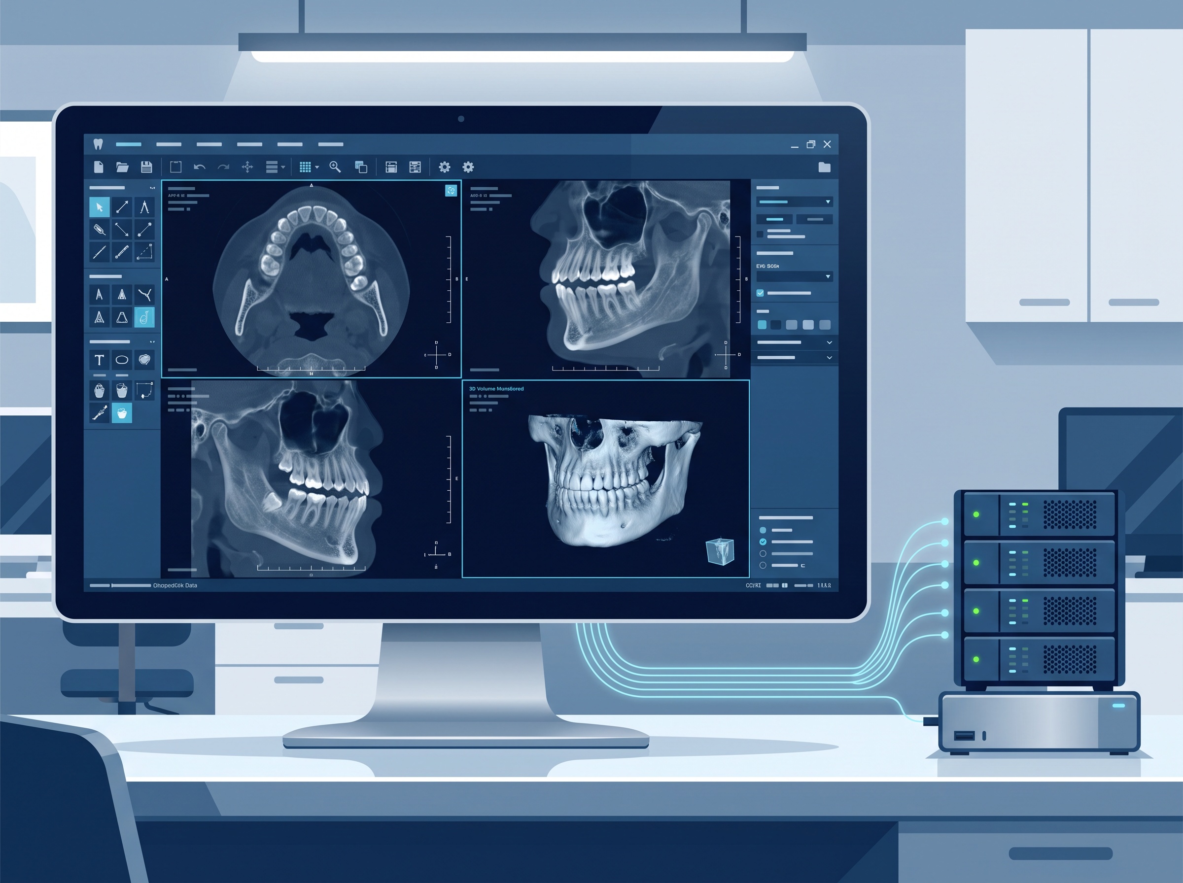
Here is where the comparison stops being purely clinical. An FMX is a handful of small files that slot neatly into your practice software. A CBCT is a large DICOM volume, and volume is the operative word – a single scan can be tens to hundreds of megabytes, and a busy practice accumulates them quickly. That has three downstream consequences the front of the office rarely sees.
Storage and backup. Large imaging files change your capacity planning and your backup windows. Records retention rules mean these scans have to survive for years – in Ontario, well beyond the visit that produced them – so they belong in a backup regime you have actually tested restoring from, not just a drive in the server closet. Imaging is a data-management discipline as much as a clinical one, a shift we traced in how dental X-ray imaging became an IT discipline.
The viewer is software, and software is attack surface. CBCT volumes are opened in dedicated DICOM viewers, and those viewers are ordinary applications that need patching like anything else. A crafted image file opening in an unpatched viewer is a real risk, not a hypothetical – as the RadiAnt DICOM advisory (CVE-2026-17264) made plain. The bigger and more specialized your imaging stack, the more of it has to be inventoried, updated, and access-controlled.
Movement and sharing. Because CBCT data is bulky and often shared with specialists, it moves – on portals, drives, and links. Every one of those hops is a place PHI can leak if it is not handled deliberately.
When to choose FMX, and when CBCT earns its dose
Choose the full-mouth series for the work general dentistry does every day: caries detection, periodontal and bone-level assessment, evaluating existing restorations, and establishing a comprehensive baseline record. For these questions FMX is not a compromise – it is the correct, lower-dose, lower-data answer, and reaching for 3D would add radiation and storage without adding diagnostic value.
Choose CBCT when the case genuinely needs three dimensions and a flat image would leave you guessing: placing an implant where bone width and vital structures must be measured, mapping an impacted tooth against a nerve, working through unusual root-canal anatomy, or assessing pathology, trauma, TMJ, and airway. In those situations CBCT honestly earns its higher dose and larger footprint, because the alternative is operating with incomplete information. The skill is not preferring one modality over the other – it is matching the exam to the question, every time.
FAQ
Is CBCT a replacement for a full-mouth series? No. They answer different questions. CBCT is for cases that need 3D detail; the FMX remains the standard for routine 2D caries and periodontal imaging. Most practices use both, for different purposes.
Does CBCT expose the patient to a lot more radiation? Usually more than an FMX, but by how much depends heavily on the field of view and machine settings – a small field-of-view scan can be close to FMX range, while a large field-of-view scan is several times higher. All of it remains far below a medical CT and within the range of ordinary background exposure. The point is to justify each scan and use the smallest field of view that answers the question.
Can we just take a CBCT on everyone to be thorough? No – that is the reasoning ALARA and image-selection criteria exist to prevent. You image in 3D when the case calls for it, not routinely.
Why does the imaging choice matter to our IT and compliance? Because CBCT produces large DICOM files that must be stored, backed up, retained for years, opened in software that needs patching, and shared securely. The modality you choose quietly shapes your storage, backup, and PHI-handling obligations.

Where Compudent fits
Choosing between an FMX and a CBCT is a clinical call – but everything that happens to the image afterward is an IT one. Compudent Systems helps dental practices across the GTA and Ontario build imaging environments that hold up: right-sized storage, tested backups, patched and access-controlled DICOM viewers, and PHIPA-conscious handling of the scans your team creates every day. If your imaging has outgrown the setup it started on, contact Compudent for an assessment – so the only thing you have to weigh in the operatory is the clinical question in front of you.
Sources & further reading:
- Full-mouth series x-rays vs. cone-beam computed tomography: What dental hygienists need to know – RDH Magazine
- ADA – Dental Radiographic Examinations: Recommendations for Patient Selection and Limiting Radiation Exposure
Related Reading
- One Screen for Scans and X-Rays: What Medit’s AuraVue With Overjet AI Means for Your Dental Practice
- The New Imaging Is Bigger Than Your Server: What Dental MRI and AI-Cleared Modalities Demand From Your Practice IT
- From Roentgen’s Lab to Your Practice Server: How Dental X-ray Imaging Became an IT Discipline