



August 5, 2026 From Roentgen’s Lab to Your Practice Server: How Dental X-ray Imaging Became an IT Discipline
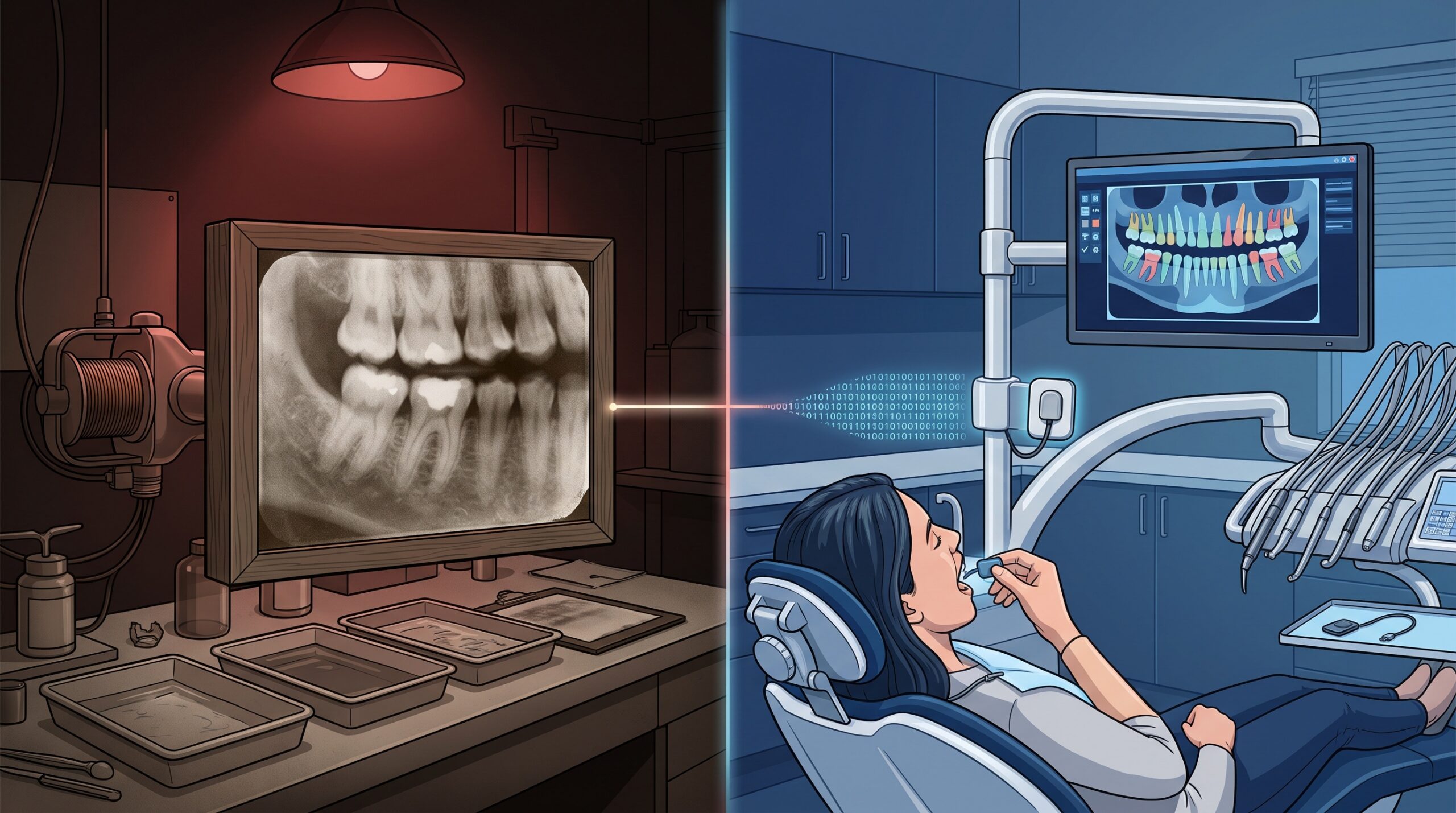
On the evening of November 8, 1895, a German physicist named Wilhelm Conrad Roentgen noticed a faint glow he could not explain and, within days, produced an image of the bones inside a human hand. He called the unknown rays X. Dentistry was among the very first fields to seize on the discovery. For most of the next century, a dental X-ray was a beautifully simple thing: a tube, a burst of radiation, a film, some chemicals, and a picture you clipped into a paper chart. The whole system had, from an IT point of view, exactly zero moving parts.
That machine in your operatory today only looks like its ancestor. Underneath, it has quietly become one of the most demanding computing systems in your entire practice.
From a darkroom to a data pipeline
The shift happened in stages, and each one added a layer of technology that someone now has to manage. Film gave way to phosphor plates and then to solid-state digital sensors that plug straight into a computer. The standalone panoramic unit grew a network port. Cone beam CT arrived and started producing not a single image but a three-dimensional volume made of hundreds of slices. Somewhere in that progression, the question stopped being “is the picture sharp?” and became “where does the picture go, who can see it, and what happens if the server holding it dies?”
In other words, imaging stopped being a clinical device you operate and became an IT system you run. Most practices crossed that line years ago without ever deciding to. The equipment vendor sold a sensor; the sensor needed a workstation; the workstation needed a server; the server needed a network, a backup, and a security posture. Nobody framed it as an infrastructure project, but that is exactly what it is.
DICOM: the language your images speak
The thing that makes modern imaging hang together is a standard called DICOM, short for Digital Imaging and Communications in Medicine. It is the reason an intraoral sensor from one manufacturer, a panoramic unit from another, and a CBCT scanner from a third can all write files that your imaging software can open, measure, and store. A DICOM file is not just a picture. It carries a wrapper of metadata around the pixels: the patient identifier, the study date, the device, the exposure settings, and more.
That is enormously powerful, and it is also a quiet responsibility. Because a DICOM file embeds patient-identifying information alongside the image, every one of those files is a small package of protected health information. Standardization made imaging interoperable, but it also means the identity of your patient travels with the picture wherever it goes. When you understand that, the reason imaging needs to be treated as an IT discipline stops being abstract.

PACS and the imaging server: where your radiographs actually live
Ask most practice owners where their X-rays are kept and the honest answer is a shrug in the direction of the back office. The technical answer is a PACS, a Picture Archiving and Communication System, or its smaller cousin, a dedicated imaging server running inside your practice management ecosystem. This is the machine that receives every image the moment it is captured, stores it, and serves it back to every operatory that asks for it.
It is also, in most practices, a single point of failure that nobody has stress-tested. If that server’s drive fails on a Tuesday morning, you have not lost a file. You have potentially lost every radiograph you have ever taken, along with the ability to work up new patients until it is restored. The imaging server deserves the same seriousness you would give the machine that runs payroll, because clinically and legally it is doing something far more consequential.
The backup math nobody does until it is too late
Here is where imaging quietly breaks the casual backup plans that practices lean on. Radiographic data does not grow gently. A busy office generates intraoral series constantly, and a single CBCT scan can be tens or even hundreds of megabytes. Multiply that across years of patients and the imaging archive becomes, by a wide margin, the largest and fastest-growing pile of data you own.
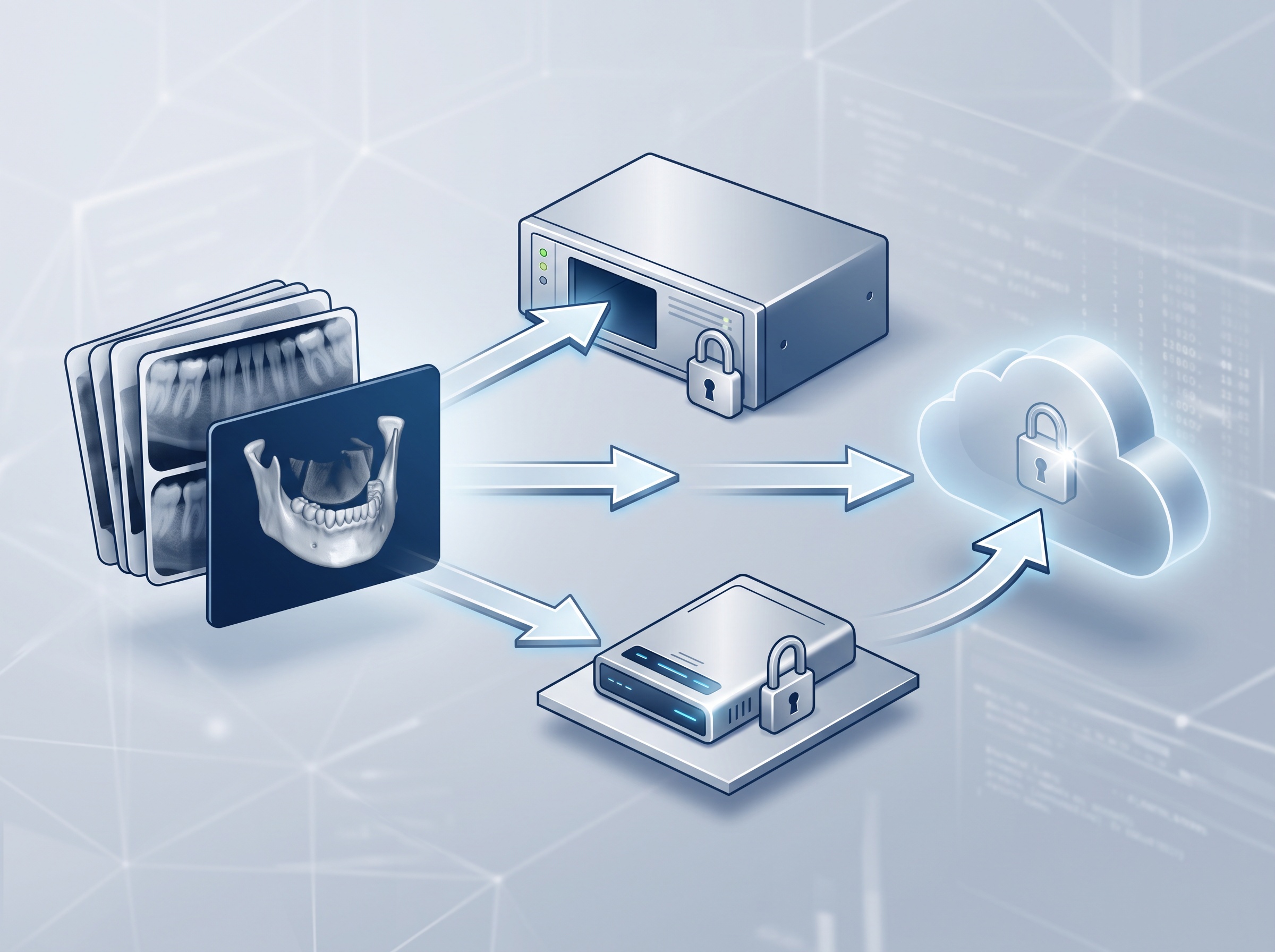
The discipline that answers this is old and boring and works: the 3-2-1 rule. Keep three copies of your data, on two different types of media, with at least one copy stored offsite. In practice that means your live imaging server, a separate local backup, and an encrypted offsite or cloud copy that leaves the building. The failure mode we see again and again is a practice that technically has a backup, but it is a single external drive sitting on the same shelf as the server, unencrypted and untested. A fire, a flood, a theft, or a ransomware event takes both at once. A backup you have never restored from is a hope, not a plan.
Your images are protected health information
It is tempting to think of PHI as the stuff in the chart, the names and treatment notes and billing. A radiograph feels different, more like a photograph than a record. Legally and practically, it is not different at all. In Ontario, patient images fall squarely under the Personal Health Information Protection Act, and the obligations that come with PHIPA apply to that imaging archive just as firmly as they apply to your clinical notes.
Concretely, that means imaging data should be encrypted both where it sits and when it moves, especially if any of it is synced to the cloud. It means access should be controlled, so that the front desk, the hygienist, and the associate each see only what their role requires, rather than everyone sharing one login to an open folder. And it means you should be able to answer the question every regulator eventually asks after an incident: who accessed this patient’s images, and when? An audit trail is not bureaucracy. It is the difference between a contained event and a reportable breach.

Uptime is now a clinical issue
When X-rays lived in a filing cabinet, the imaging system never went down. The cabinet did not crash, get encrypted, or lose network connectivity. The tradeoff of digital imaging is speed and diagnostic power in exchange for dependence on systems that can fail. When the imaging server is unreachable, you cannot pull prior films, you cannot capture new ones into the record, and the schedule backs up behind a problem that has nothing to do with dentistry.
Treating imaging as an IT discipline means planning for that day before it arrives: redundancy so a single component failure does not stop the practice, monitoring so problems are caught before they become outages, and a recovery plan that has actually been tested against the clock. Hardware will eventually fail. The real question is how long your practice is down when it does, and whether you decided that number in advance or found it out the hard way.

One long thread, from 1895 to your server room
The story of the dental X-ray is one continuous thread: a glowing discovery in a Wurzburg laboratory, a century of film and chemistry, and a present in which every image is a data object that must be captured, standardized, stored, secured, backed up, and served without interruption. Roentgen gave us the picture. What we have built around it since is, unmistakably, information technology, and the practices that thrive with modern imaging are the ones that recognized this and resourced it accordingly.
Compudent Systems designs, secures, and supports the imaging and IT infrastructure that GTA and Ontario dental practices depend on every day, from DICOM-aware imaging servers and 3-2-1 backup strategies to PHIPA-aligned security, monitoring, and tested recovery plans. If you are not certain how your radiographs are stored, how quickly you could recover them, or who can see them, that is the conversation worth having now rather than after an outage. Contact Compudent Systems for an assessment of your practice’s imaging infrastructure and data protection.
Sources & further reading:
- DICOM – Digital Imaging and Communications in Medicine (official standard site)
- Personal Health Information Protection Act, 2004 (PHIPA), Ontario
The Story of the X-Ray, Across Our Network
How dental X-ray imaging became an IT discipline — the fuller history behind it:
- How Marie Curie drove X-ray trucks into the First World War
- What your dentist actually sees on an X-ray, 130 years on
- The equipment history from gas tubes to digital sensors
Related Reading
- One Screen for Scans and X-Rays: What Medit’s AuraVue With Overjet AI Means for Your Dental Practice
- The New Imaging Is Bigger Than Your Server: What Dental MRI and AI-Cleared Modalities Demand From Your Practice IT
- You Can’t Patch That Old Sensor or CBCT: How to Safely Keep Legacy Imaging Devices on Your Dental Network