



September 4, 2026 The New Imaging Is Bigger Than Your Server: What Dental MRI and AI-Cleared Modalities Demand From Your Practice IT
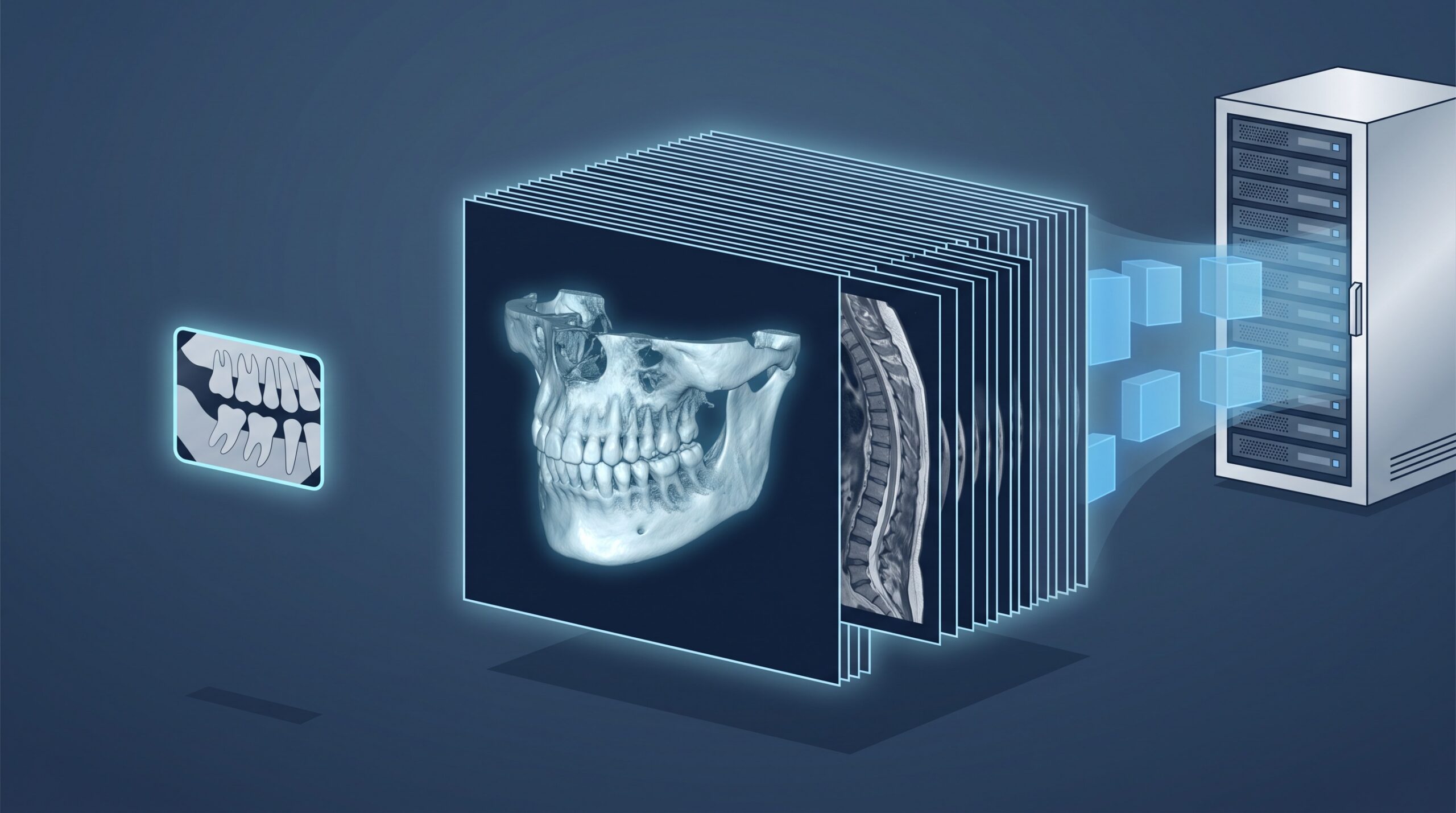
The clinical headlines are genuinely exciting for once. In 2026 the FDA cleared the first dental-dedicated MRI system – Dentsply Sirona and Siemens Healthineers’ MAGNETOM Free.Max Dental Edition – bringing radiation-free, soft-tissue imaging into the dental office, and it arrived alongside a run of new 3D and AI-assisted clearances that one trade headline summed up as four FDA clearances reshaping dentistry. The clinical press has that story well covered. There is a second story it mostly doesn’t tell, and it is the one that lands on your desk after the equipment rep leaves: every one of these advances produces a much bigger, richer image, and that image has to be captured, moved, viewed, stored, retained, and backed up on infrastructure most practices sized years ago for digital radiographs. The new imaging is, quite literally, bigger than a lot of practice servers were built to hold.
What actually got cleared – and why it’s a trend, not a gadget
It helps to see this as a wave rather than a single product. The dental-dedicated MRI is the marquee item because it brings an entirely new, radiation-free modality to dentistry for soft-tissue and temporomandibular imaging. But it is landing at the same time as new FDA-cleared 3D imaging and AI-assisted detection tools that read radiographs and CBCT volumes for the clinician. The common thread across all of them is data density: MRI produces multi-image series, modern 3D units produce ever-higher-resolution volumes, and AI tools generate additional annotated derivatives of each study. None of this replaces your existing CBCT and sensors – it stacks on top of them. So the practical question for a practice isn’t “MRI, yes or no?” It is “is our infrastructure ready for imaging that is several times heavier than what we run today?” And that question deserves the same rigour as choosing which imaging modality your practice actually needs and when.

Why bigger images are an IT problem, not just a clinical win
A single 2D bitewing is a small file. A high-resolution CBCT volume is dramatically larger; an MRI series larger still; and an AI-annotated study adds yet another layer on top of the original. Individually, none of these is alarming – modern storage is cheap. The trap is the curve. Take a bigger average study size, multiply it by the number of studies your practice captures every day, and compound that across the years of retention a dental record requires, and you get an accumulation curve that climbs far faster than the gentle slope film-replacement radiography produced. Practices that sized their storage, network, and backups for 2D imaging and never revisited the assumption are the ones that get surprised – not by the first MRI scan, but by month eighteen, when the drive that was “plenty” is suddenly at ninety percent and the nightly backup no longer finishes before the team arrives.

The server and storage question
Start with where the images actually live. Many practices keep imaging on a local server or an imaging workstation acting as one, and that is perfectly workable – provided the capacity is planned against the new curve, not last year’s. The right conversation covers headroom (size for two to three years of growth, not this month’s footprint), architecture (local storage versus a proper PACS versus a hybrid that tiers older studies to cloud), and retention. Retention is not optional: under PHIPA and HIPAA, diagnostic images are part of the patient record and must be kept for years, so “we’ll delete old scans to make room” is not a storage strategy – it is a compliance problem. The goal is a storage plan that expects the growth, tiers it sensibly, and never forces a clinical-versus-capacity trade-off at the front desk.
The network and the workstation that has to open it
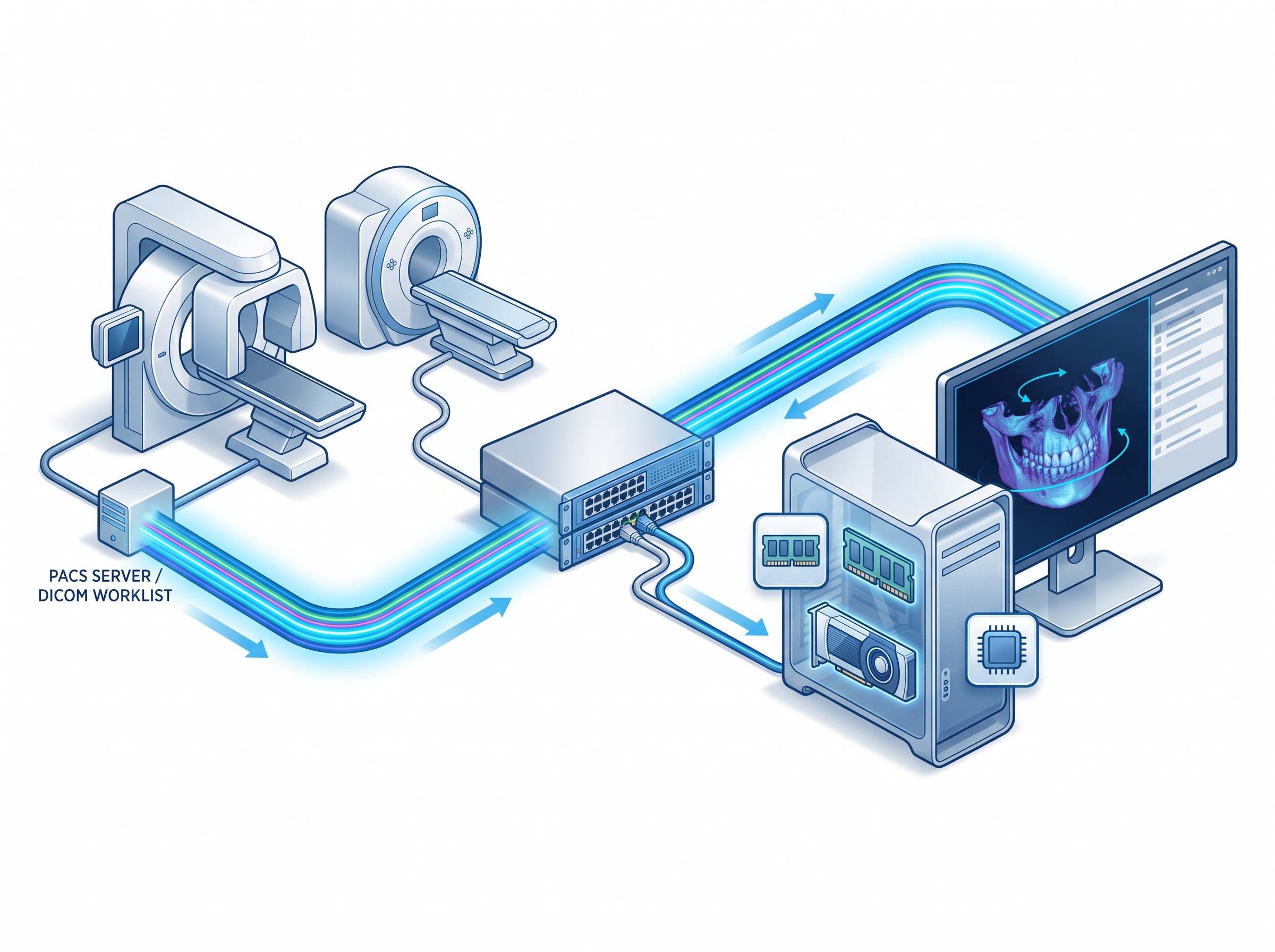
Big images have to travel, and then someone has to open them. On the network side, moving 3D volumes and MRI series between the modality, the server, and the operatory where the dentist reviews them puts real load on office cabling and switches that were fine when the heaviest traffic was a panoramic. On the workstation side, a review PC needs enough RAM and graphics capability to load and manipulate a large volume smoothly – a dentist rotating a CBCT or scrolling an MRI stack on an underpowered machine simply waits, and waiting at the chair is expensive. Underneath both sits the plumbing: the DICOM worklist and PACS pathways that route a study from capture to the right patient chart. When those are healthy the workflow is invisible; when they are undersized, every heavy new modality makes the stalls more obvious. The same data-flow discipline is exactly what makes AI imaging tools usable in practice, which is why integrating an AI platform like Overjet is as much an integration and data-governance project as a clinical one.
Backups and business continuity – the images are the record
Here is the part that is easy to skip and painful to relearn: the images are not a by-product of care, they are the clinical record, and larger datasets make protecting them both more important and more demanding. Bigger studies stretch backup windows – the nightly job that comfortably finished at 2 a.m. can start bleeding into the working day – and they raise the cost of whatever off-site or cloud copy you keep. That is not a reason to back up less; it is a reason to design the backup deliberately: a genuine 3-2-1 approach (multiple copies, more than one medium, at least one off-site and ideally immutable), sized for the new data volume, and – the step everyone forgets – tested restores, because a backup you have never restored is a hope, not a safeguard. This is also where new imaging meets old equipment on the same network. As you add heavier modern modalities, the aging units that cannot be updated still need containment, which is a discipline in its own right: keeping unpatchable legacy imaging devices safely segmented so the growing, valuable image store they sit beside stays protected.

The takeaway: size the IT during procurement, not after install
The single most useful habit a practice can adopt is to treat an imaging purchase as an IT project, not just a clinical one. Before the unit is ordered – not after it is bolted to the wall – ask what average study size it produces, how much daily volume that adds, whether the server has the headroom and the retention plan to absorb years of it, whether the network and review workstations can move and open the images without stalling, and how the backup window and cost change. None of these are reasons to hesitate on adopting better imaging; the diagnostic gains from radiation-free MRI and high-resolution 3D are real. They are simply the questions that separate a smooth rollout from a scramble six months later. Plan the infrastructure with the same care you plan the clinical use, and the new imaging becomes an upgrade instead of a surprise.
If your practice is weighing a new imaging modality – a dental MRI, a next-generation CBCT, or an AI platform layered on top of what you already run – and you want to know whether your server, network, and backups are ready for it, contact Compudent Systems. We help dental practices across Ontario size storage and networks for modern imaging, design PACS and backup strategies that meet PHIPA retention obligations, and make sure the infrastructure is ready before the equipment arrives – so the only surprise is how good the images look.
Sources & further reading:
- From the first dental MRI to orthodontic AI: Four FDA clearances reshaping dentistry – Oral Health Group
- Dentsply Sirona Advances Dental Imaging With MRI – Dentistry Today
- First-ever Dental Dedicated MRI Validated in Clinical Trial and Cleared by FDA for Advanced Soft-Tissue Diagnostics – Dentsply Sirona