



October 13, 2026 Dental MRI Is Arriving in Practices — and It Is an IT Project Before It Is a Clinical One
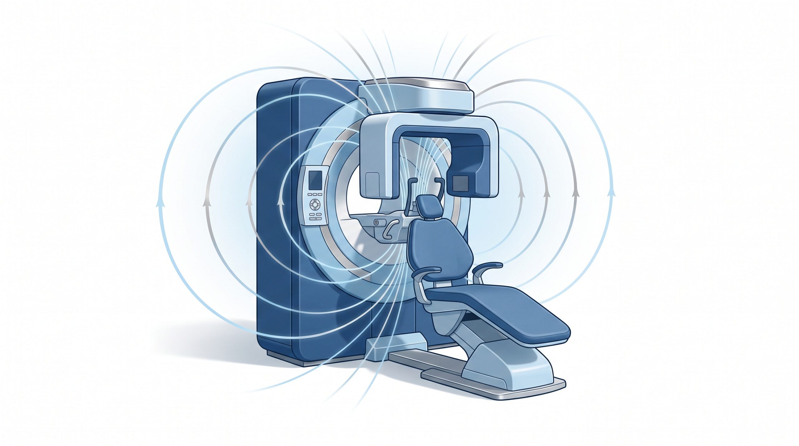
For more than a century, the dental image has been a picture made with radiation — film, then phosphor plates, then digital sensors, then the 3D volumes of cone-beam CT. A new modality is now entering the operatory that breaks that pattern entirely: magnetic resonance imaging. In early 2026 the first dental-dedicated MRI (ddMRI) system cleared the U.S. FDA, and it has since begun reaching practices through the major dental distributors. It uses no ionizing radiation at all, and it is built to see the soft tissue that X-ray and CBCT render poorly — pulp, nerves, the temporomandibular joint, and soft-tissue pathology.
The clinical case will be made at every study club and trade show this year. This article is about the part that will not be on the booth banner: a dental MRI is an IT project before it is a clinical one. The scan is radiation-free, but the data it produces is anything but weightless, and the network, storage, backup, and integration it demands land squarely on your practice’s technology — the same way every previous imaging advance eventually did.
What the new modality actually is
The system that received clearance is a low-field, helium-free MRI designed specifically for the head and jaw rather than the whole body. The low-field, helium-free design matters because it lowers the siting and facilities burden that has always kept MRI in hospitals and imaging centres — less specialized shielding and infrastructure than a conventional high-field scanner. It is still a magnet, and siting still deserves professional planning, but the premise is a scanner that can realistically live in a dental practice. For the clinician, the draw is straightforward: a radiation-free way to image structures that current dental imaging shows only indirectly.
This continues a long arc we have written about before — the steady transformation of dental imaging from a darkroom craft into a data discipline. If MRI feels like a leap, it is worth remembering that every modality felt that way once; our piece on how dental X-ray imaging became an IT discipline traces how each generation of technology quietly moved the work from the operatory to the server room. MRI is the biggest step in that direction yet.
The clinical leap comes with a data leap
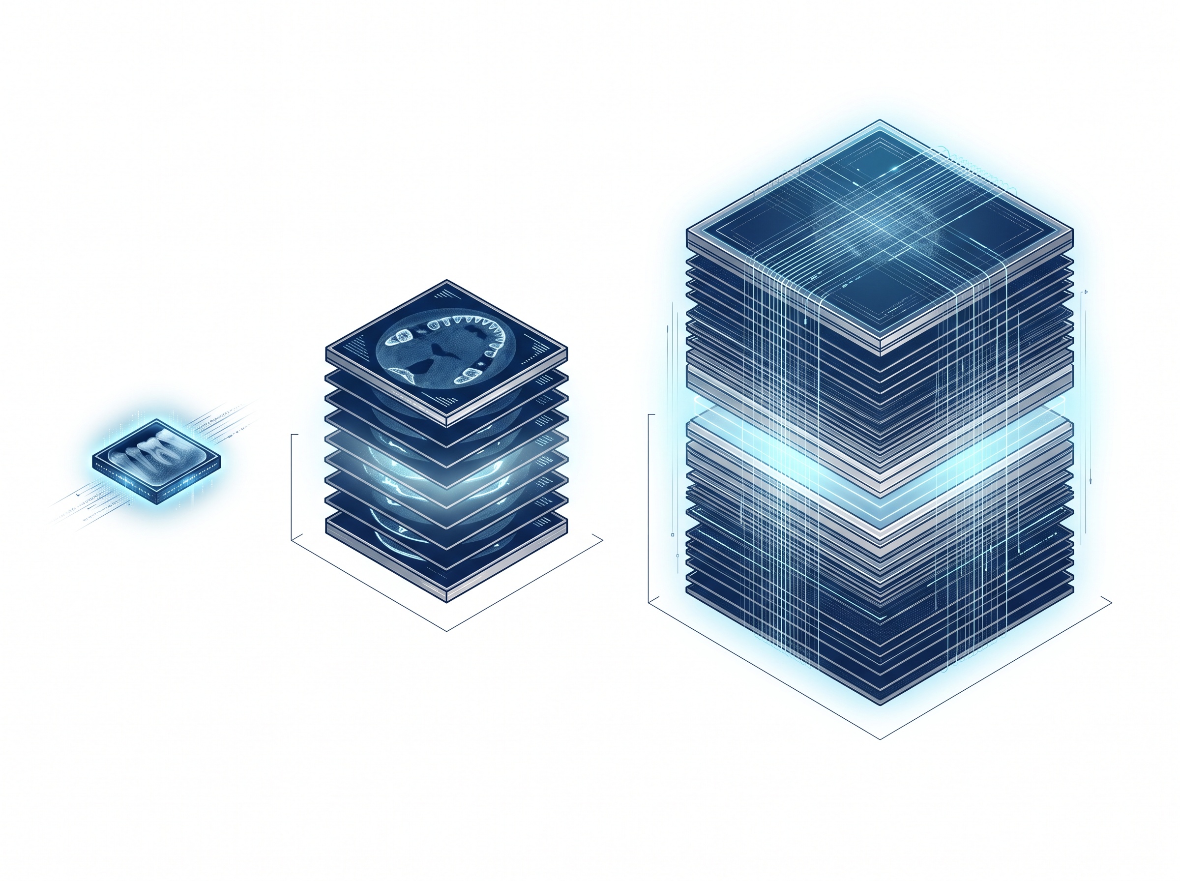
Here is the number that should frame every planning conversation: an MRI study is large. A single intraoral radiograph is a modest file. A CBCT volume is substantially bigger, and most practices already felt that jump when they adopted 3D. A volumetric MRI dataset is larger still — and unlike a quick bitewing, it is the kind of study a practice will want to keep, compare over time, and retain for the full records-retention period.
Multiply one large study by a schedule of patients, across the years your regulatory obligations require you to keep records, and the storage question stops being an afterthought. This is not a reason to avoid the technology; it is a reason to size for it honestly. A practice that bolts a high-volume modality onto storage that was specified for intraoral images and a bit of CBCT will run out of room, slow down, or quietly stop backing everything up — usually discovering which, at the worst possible moment.
It has to move, land, and be found
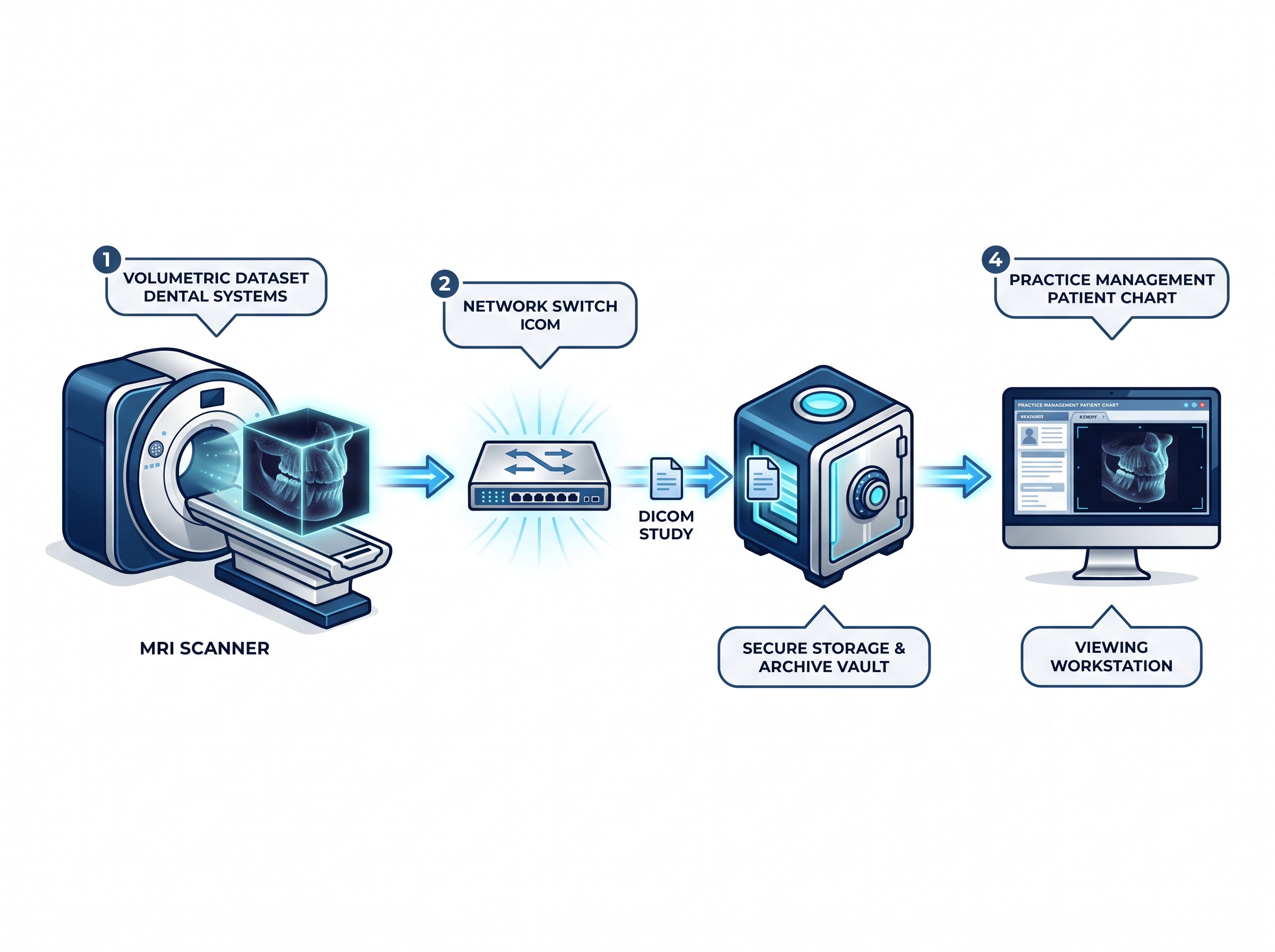
Capturing the image is only the first step. Every study then has to travel your network, land in storage, and file itself into the correct patient record where a clinician can actually open it. Large datasets moving across an office network that was sized for email and intraoral images will expose every weak link — an aging switch, a tired wireless segment, a workstation that cannot load a big volume without stalling.
Then there is integration, which is where many imaging headaches actually live. A new modality has to speak to your practice management system through the DICOM standard and the connectors — the imaging bridges — that tie acquisition to the patient chart. We have written at length about how fragile that layer can be in our explainer on imaging bridge software; the lesson applies doubly to a brand-new modality. Before you buy, the question is not “does it make great images” but “is there a tested path for those images into the system my team already uses every day, attached to the right patient, viewable on the workstations we have?”
Backup and retention are not optional extras
Larger, clinically valuable studies raise the stakes on the least glamorous part of IT: backup. If a radiograph is a diagnostic record you are legally required to retain, an MRI study is that same obligation at many times the size. Your backup and archive strategy has to absorb the new data volume without silently falling behind, and it has to be verifiably restorable — not merely “running.”
This is also where imaging intersects with security. Valuable, hard-to-recreate patient data is exactly what ransomware operators hold hostage, which is why we keep returning to the theme of a backup you actually control — immutable and air-gapped. Adding a modality that generates large, irreplaceable studies makes that discipline more important, not less. A practice that cannot restore its imaging archive has not bought an imaging upgrade; it has bought a bigger single point of failure.
A scanner is also a networked computer
A dental MRI, like every modern imaging device, is a networked computer running an operating system, sitting on your practice network, often with vendor remote-access for support. That makes it part of your attack surface as much as your clinical arsenal. It deserves the same discipline as any connected medical device: placed on a segmented part of the network, kept patched or otherwise protected where it cannot be, and covered by a clear understanding of who is responsible for its software security over its service life. The clinical conversation is about what the magnet can see; the IT conversation is about making sure the device cannot become the way an attacker sees everything else.

The questions to ask before the purchase order
You do not need to be an imaging engineer to protect the investment — you need to force the infrastructure questions to the surface while you still have leverage, the same discipline the latest imaging guidance on selecting and documenting studies brings to the clinical side.
- How much data per study, and how much per year? Get a concrete figure from the vendor and size storage and backup for several years of it, not for launch week.
- Is there a tested DICOM path into our specific practice management system? Ask for written confirmation and, ideally, a demonstration with your software — not “it supports DICOM” in the abstract.
- Will our network and viewing workstations actually handle these studies? Have someone assess the switches, cabling, and the computers clinicians will use to open large volumes, before install day.
- Can we back up and verifiably restore the new data? Confirm the archive strategy absorbs the volume and that a restore has actually been tested.
- How is the device secured and supported over its life? Clarify patching, remote-access, and network placement, and who owns each.
The takeaway
Dental MRI is a real clinical advance and a genuinely exciting one — a radiation-free window onto tissue that dentistry has long struggled to image. But excitement is not an implementation plan. The practices that adopt it well will be the ones that treated it, from the first conversation, as the infrastructure project it quietly is: sizing storage, hardening the network, proving the integration, verifying the backups, and securing the device. The magnet is the easy part to admire. The data is the part that has to be engineered.
How Compudent Systems can help
At Compudent Systems, we help dental practices across the GTA and Ontario bring new imaging technology online without the hidden surprises — sizing the storage and backup, checking that the network and workstations can carry the load, confirming a real integration path into your practice management system, and securing connected imaging devices as the networked computers they are. If a dental MRI, a new CBCT, or any imaging upgrade is on your horizon, contact Compudent Systems for an imaging-readiness assessment, and make sure the infrastructure is ready before the equipment arrives.
Sources & further reading:
Related Reading
- The New Imaging Is Bigger Than Your Server: What Dental MRI and AI-Cleared Modalities Demand From Your Practice IT
- One Screen for Scans and X-Rays: What Medit’s AuraVue With Overjet AI Means for Your Dental Practice
- How Dental AI Tools Connect to Your Imaging Software: TWAIN, DICOM, API, and PMS Bridges