




November 13, 2026 The 2026 ADA Dental Radiograph Guidelines Are Here: How Often to Take X-Rays, the New CBCT Rules, and What Your Practice Must Document
For more than a decade, the reference point for who should receive dental x-rays and how often was the 2012 ADA and U.S. FDA document, “Dental Radiographic Examinations: Recommendations for Patient Selection and Limiting Radiation Exposure.” In 2026 that guidance was finally brought up to date: the American Dental Association, together with the American Academy of Oral and Maxillofacial Radiology (AAOMR), co-published updated consensus recommendations covering both conventional planar (2-D) imaging and cone-beam computed tomography (CBCT, 3-D). It is the most significant refresh of dental imaging selection guidance in years — and while the headlines are clinical, the update lands squarely on the records, software, and compliance side of a practice too.
Here is what the 2026 guidance actually says in practical terms, and what it means for the way your practice documents, stores, and defends every image it captures.
There is still no fixed x-ray schedule — and that is the whole point
The single most misunderstood thing about dental radiograph guidance is the expectation of a timetable: x-rays every six months, a full series every so many years. The guidance has never worked that way, and the 2026 update reaffirms it. Imaging is ordered on the basis of patient-selection criteria — the individual patient’s history, clinical examination, signs, symptoms, and risk — not on a calendar and emphatically not on what a dental insurance contract happens to allow. A radiograph is justified when it is expected to change diagnosis or treatment, and not before.
That principle sits on top of ALARA — keeping radiation exposure As Low As Reasonably Achievable. Every exposure should be the minimum needed to answer a real clinical question. The guidance exists precisely so that imaging is driven by need rather than habit.
The one exam with a frequency recommendation: the bitewing
If patients ask you to put a number on it, the honest answer is that the bitewing is effectively the only examination the guidance attaches an interval to — and even that interval is risk-based rather than fixed. In broad strokes, a recall patient with clinical caries or other increased risk factors may warrant posterior bitewings at roughly 6 to 12 month intervals, while a recall patient with no clinical caries and no increased risk can go substantially longer — on the order of 18 to 36 months for adults, with shorter intervals for children because their teeth and caries risk change faster. The exact interval is a clinical judgment the dentist makes and records; the guideline gives the framework, not a stamp.
Everything else — panoramic imaging, full-mouth series, and especially 3-D imaging — is ordered when the clinical situation calls for it, not on a timer.
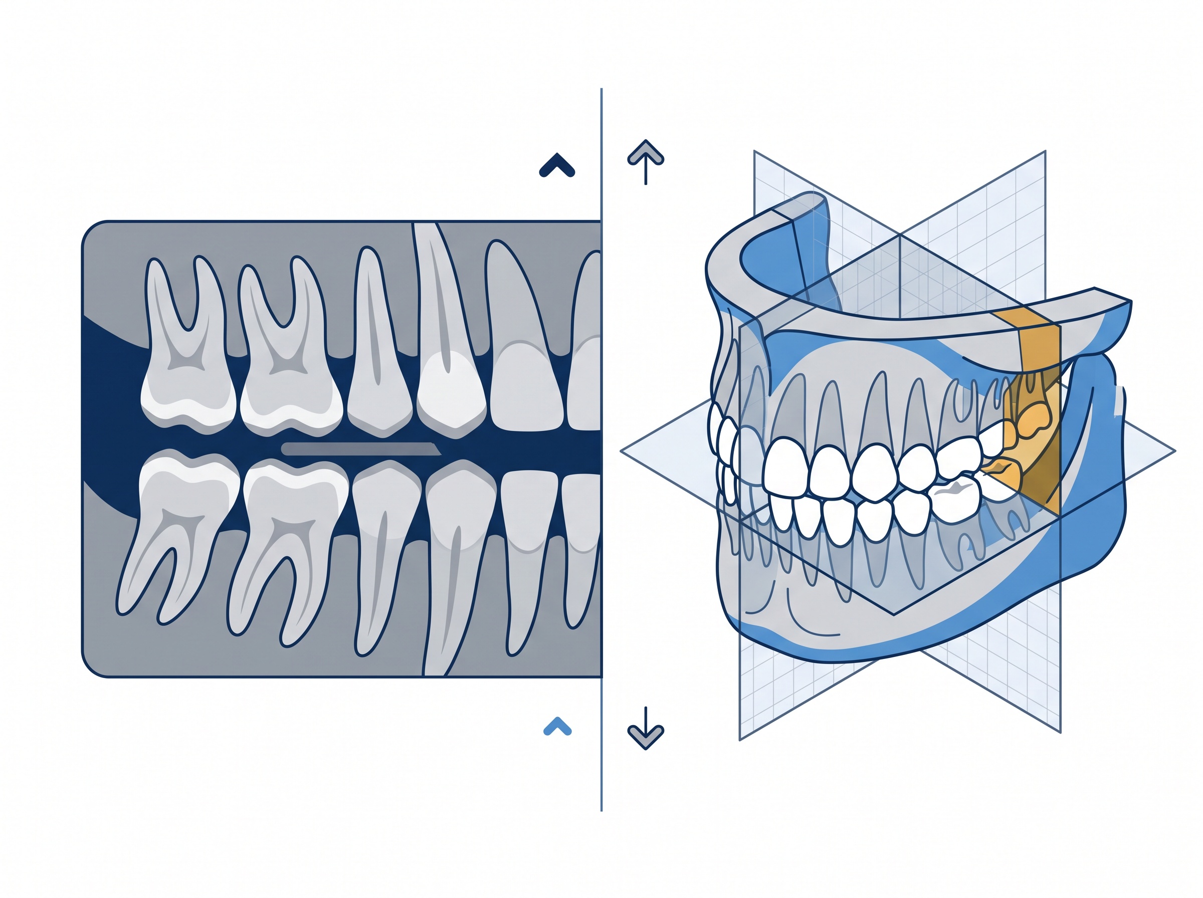
The real headline: CBCT finally gets its own rules
The most consequential change in 2026 is the explicit, scenario-based treatment of cone-beam CT. The 2012 document predated the routine presence of CBCT in general practice; the new AAOMR-backed recommendations address specific clinical situations in which 3-D imaging is — and is not — justified, from implant planning to certain endodontic and surgical scenarios. The throughline is dose. CBCT delivers far more diagnostic information than a 2-D film, but at a higher radiation cost, so the guidance pushes clinicians to reach for it deliberately rather than reflexively, and to prefer the lowest-dose modality that can still answer the question.
For a practice that has invested in a CBCT unit, this matters operationally as well as clinically: a scan is a large, information-dense study that has to be stored, protected, and defended as a reasoned clinical decision. Which brings the whole update back to something that is very much your IT and records responsibility.
A selection criterion you cannot prove is a selection criterion you did not follow
Here is the part that is easy to miss. “Imaging is justified by clinical need” is not only a clinical statement — it is a documentation requirement. If every exposure is supposed to be driven by a specific indication and the ALARA principle, then the practice has to be able to show, for any given radiograph, why it was taken: the clinical indication, the risk assessment, and the judgment that no lower-dose alternative would do. If that reasoning is not captured in the patient’s record, the practice has, in effect, no evidence that it followed the guidance at all.
This is where the imaging software and practice-management system earn their keep. The guideline is only as good as the record that backs it, and that record has to be captured at the moment of care, stored with the image, and retrievable later. The same discipline we described around keeping clinical conversations and PHI inside compliant systems applies here — see our guidance on what HIPAA and PHIPA actually require when a practice handles patient information. A diagnostic image with no documented justification is a liability hiding in your archive.

Ontario practices: the ADA criteria sit on top of HARP, not instead of it
For practices in Ontario and across Canada, it is worth being precise about how these pieces fit together. The ADA/AAOMR recommendations are clinical patient-selection guidance — the “when and why” of imaging. They do not replace the legal radiation-safety regime, which in Ontario is the Healing Arts Radiation Protection (HARP) Act: equipment registration, inspection, shielding, and operator requirements that govern the machine and the room. The two operate together. The HARP Act makes sure the x-ray source is safe and compliant; the ADA selection criteria make sure each exposure is clinically warranted in the first place. If you are unclear on the equipment-safety side, our explainer on what a dental x-ray inspection actually checks under the HARP Act covers it in detail. Following the ADA guidance does not exempt a practice from HARP, and vice versa.
Retention, backup, and access: images are part of the record for years
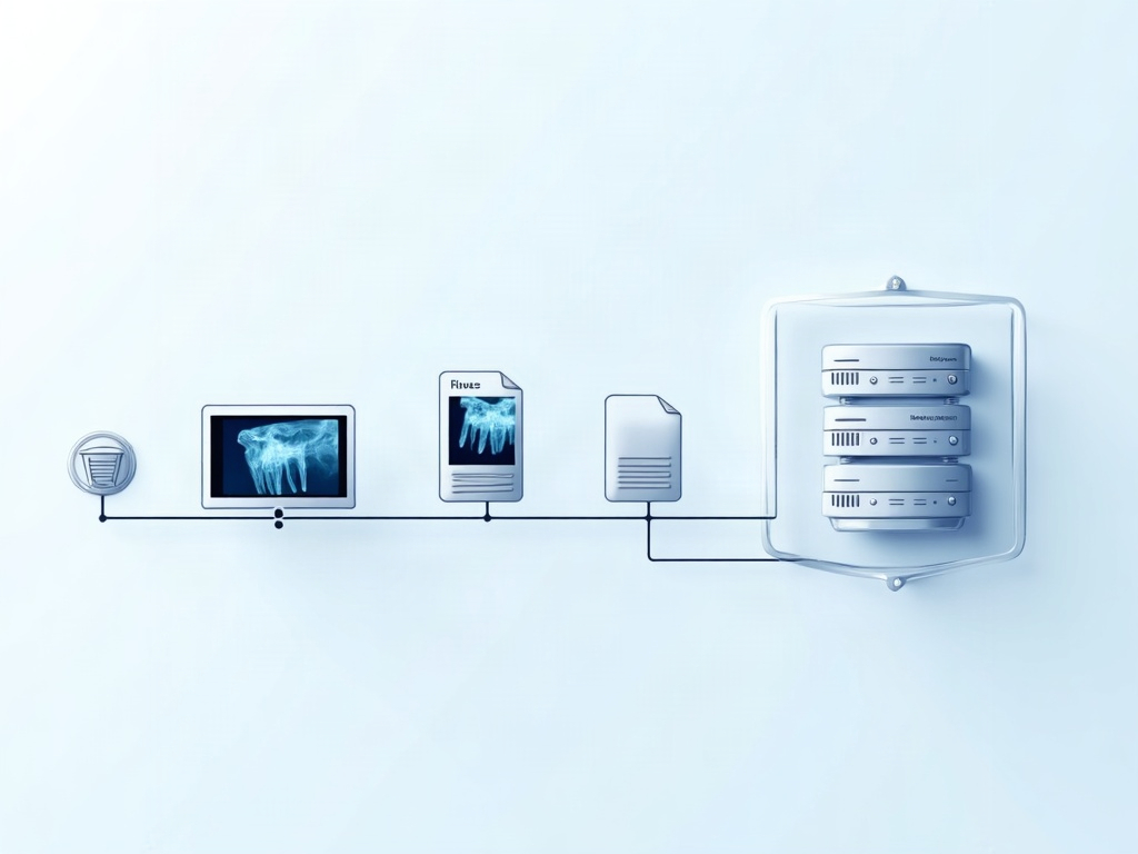
A radiograph is not a disposable snapshot — it is part of the clinical and legal record, subject to retention obligations that run for years and, for images taken of minors, well into adulthood. That has concrete infrastructure consequences. The images have to be stored where they will survive hardware failure, kept backed up in a way that is itself protected against ransomware, and remain accessible and legible for the full retention period even as software and formats change. Under PHIPA, they also have to be kept secure and access-controlled the entire time. Capturing the right image for the right reason is only half the obligation; being able to produce it, intact and attributable, years later is the other half — and that is a storage, backup, and records-management problem that belongs to the practice’s IT side.
What to do with the update
The 2026 ADA/AAOMR guidance does not ask most practices to image more or less as a rule — it asks them to image deliberately, document the reason, and treat 3-D imaging with particular care. Translated into an operational checklist, that means a few things worth reviewing now: confirm your clinicians are selecting imaging on risk and indication rather than a default recall schedule; make sure your imaging software actually captures the clinical justification alongside each study; be especially disciplined about when CBCT is ordered and how those large studies are stored; and verify that your retention, backup, and access controls meet PHIPA for the full life of the record.
Most of that lower half — the documentation, storage, retention, and security of your diagnostic images — is exactly where a practice’s technology either supports the guidance or quietly undermines it. If you want your imaging software, archive, and backups aligned with the 2026 recommendations so that every image you take is both clinically defensible and properly retained, contact Compudent Systems for a review. We will help you make sure the record keeps up with the clinical standard.
Sources & further reading:
- X-Rays/Radiographs — Oral Health Topics (American Dental Association)
- The Selection of Patients for Dental Radiographic Examinations (U.S. FDA)
Related Reading
- The ADA Now Says Scan Less — So Your Records Have to Say Why: What the 2026 Imaging Guidelines Mean for Your Practice Software
- One Screen for Scans and X-Rays: What Medit’s AuraVue With Overjet AI Means for Your Dental Practice
- FMX vs. CBCT: Which Dental Imaging Does Your Practice Actually Need, and When